Medical Imaging for Detection, Diagnosis and Treatment
Radiology uses X-rays, radioactive tracers, magnetic waves and ultrasonic waves to obtain detailed images of inside the body. Doctors use these images to detect and diagnose illnesses and injuries, as well as to help develop treatment plans.
Palmdale Regional Medical Center offers a wide range of radiology services to residents of the Antelope Valley and the surrounding region, including:
- Diagnostic radiology
- Computed tomography (CT) and CT angiography (CTA)
- Magnetic resonance imaging (MRI)
- Diagnostic ultrasound
- Nuclear medicine
- Interventional radiology
For more information about the medical imaging services at Palmdale Regional Medical Center, please call 661-382-5050.
Advanced Imaging Technology
The advanced diagnostic imaging technology used at Palmdale Regional Medical Center gets results to your physician quickly, so he or she can make a diagnosis and recommend treatment for you as soon as possible. Our technology means that your physician can access your results immediately, from any location, through our secure imaging website.
X-ray
An X-ray image is produced when a small amount of radiation passes through the body to create an image on sensitive digital plates on the other side of the body. The ability of X-rays to penetrate tissues and bones depends on the tissue’s composition and mass, and the difference between these two elements creates the image. Contrast agents, such as barium, may be swallowed to outline the esophagus, stomach and intestines to help provide better images of an organ.
Computed Tomography
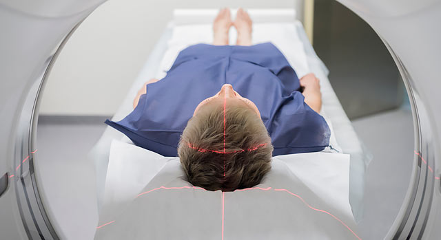
Computed tomography, also known as CT or CAT scan, generates detailed images of an organ by using an X-ray beam to take images of many thin slices of that organ and joining them together to produce a single image. The source of the X-ray beam circles around the patient and the X-rays that pass through the body are detected by an array of sensors. Information from the sensors is computer processed and displayed as an image on a screen.
Palmdale Regional Medical Center uses advanced equipment for CT examinations. Each time the scanner rotates around a patient's body, it uses low radiation X-rays to create 16 or 64 high-resolution slices (images) depending on the scanner. Because the scanner circles a patient's body about four times every second, patients may have to lie in the CT machine for two to three minutes.
Magnetic Resonance Imaging
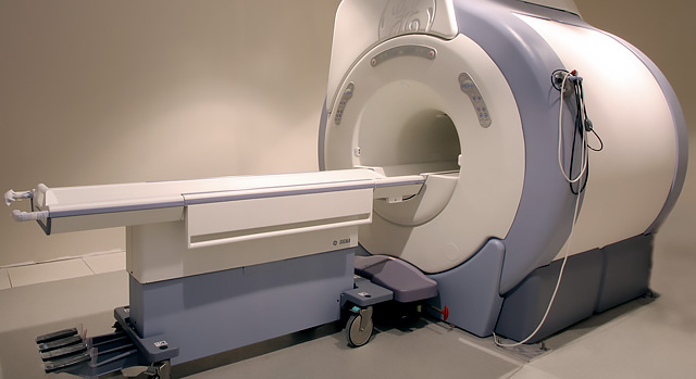
MRI uses radio waves and a strong magnetic field to create clear, detailed images of internal organs and tissues. Since X-rays are not used, no radiation exposure is involved. Instead, radio waves are directed at the body’s protons within the magnetic field. This exam takes 30-50 minutes on average and consists of several imaging series. Most studies will require a small intravenous injection of a contrast agent that usually contains the metal Gadolinium. MRI contrast does not contain iodine, an element used in other contrast agents for X-rays or CT scans, so it may be safer for patients who are sensitive to iodine.
Ultrasound (Sonography)
High frequency sound waves are used to see inside the body. A transducer, a device that acts like a microphone and speaker, is placed in contact with the body using a special gel that helps transmit the sound. As the sound waves pass through the body, echoes are produced and bounce back to the transducer.
By reading the echoes, the ultrasound can produce images that illustrate the location of a structure or abnormality, as well as provide information about its composition. Ultrasound is a painless way to examine the heart, liver, pancreas, spleen, blood vessels, breast, kidney or gallbladder, and is a crucial tool for obstetrics.
Abdominal Aorta Ultrasound
The aorta is the largest blood vessel in your body and distributes oxygenated blood from your heart throughout your circulatory system. Over time, the walls of your aorta may weaken, allowing it to balloon. When that happens and the diameter of your aorta reaches 1.18 inches or larger, it is called an abdominal aortic aneurysm (AAA).
As the diameter on an aneurysm increases, the risk that it will rupture increases as well. If your doctor suspects that you could have an aortic aneurysm, he or she may ask you to have an abdominal aorta ultrasound. This test uses high-frequency sound waves to produce an image of your aorta so that doctors can determine its size. Based on the results, your physician may suggest a procedure to repair the weakened walls, or may suggest periodic ultrasounds to monitor the size of your aorta over time to determine when additional treatment is necessary.
If you are a male age 60 and older, smoke or have smoked and have a family history of abdominal aortic aneurysm, you should talk with your physician to decided if this ultrasound screening is recommended for you.


